Understanding 3D Imaging Ultrasound: A Revolutionary Tool for Expecting Parents
Pregnancy is a transformative journey, one filled with joy, anticipation, and sometimes uncertainty. Expecting parents often rely on ultrasound technology to monitor the health of both mother and baby, providing vital information about fetal development. Among the various advancements in ultrasound technology, 3D imaging ultrasound has emerged as a revolutionary tool, offering a detailed and vivid view of the developing fetus. This article explores how 3D imaging ultrasound enhances the prenatal experience for expecting parents and the medical professionals guiding them through pregnancy.
What is 3D Imaging Ultrasound?
Ultrasound imaging uses sound waves to create images of the inside of the body, offering valuable insight into fetal development, maternal health, and potential complications. Traditional 2D ultrasound provides flat, black-and-white images of the fetus, helping to assess basic anatomical features, heart rate, and growth.
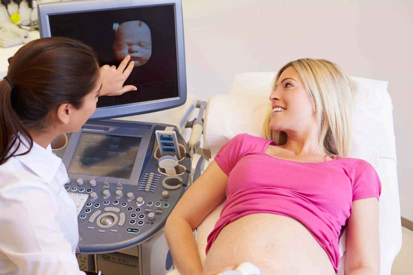
However, 3d imaging ultrasound goes a step further by capturing multiple 2D images from various angles and then processing them to create a three-dimensional representation. The result is a more lifelike, detailed image of the baby that allows medical professionals to evaluate the fetus in greater detail. This 3D technology enhances the understanding of the baby’s development, especially in the later stages of pregnancy.
The Benefits of 3D Imaging Ultrasound for Expecting Parents
1. Enhanced Visuals and Bonding Opportunities
One of the most significant advantages of 3D imaging ultrasound is its ability to provide expecting parents with a more tangible connection to their baby. Unlike traditional ultrasound images, which can be abstract and difficult to interpret, 3D imaging ultrasound allows parents to see their baby’s facial features, hand movements, and even expressions in vivid detail. This sense of realism can enhance the emotional bond between the parents and their unborn child, making the pregnancy feel more tangible and personal.
For many parents, seeing a 3D image of their baby before birth is an emotionally powerful experience. It offers a clearer understanding of the baby’s features, such as facial structure and skin tone, which can be especially meaningful for parents anticipating a healthy birth.
2. Improved Diagnostic Accuracy
While 3D imaging ultrasound is often used for bonding purposes, it also serves a critical medical function. The increased level of detail provided by 3D ultrasound allows healthcare providers to identify potential issues that may not be visible in traditional 2D imaging. For instance, doctors can more accurately detect abnormalities in the baby’s facial features, such as cleft lip or palate, or assess the development of organs and limbs.
In cases of complex pregnancies, 3D imaging ultrasound can also offer a more detailed view of the placenta, amniotic fluid, and the positioning of the fetus, helping doctors plan for safer delivery options. By improving diagnostic capabilities, this technology empowers healthcare providers to make more informed decisions about patient care.
3. Non-invasive and Safe for Both Mother and Baby
One of the main appeals of 3D imaging ultrasound is that it is non-invasive and considered safe for both the mother and the baby. It uses sound waves, which do not carry the same risks as radiation-based imaging methods like X-rays. As such, 3D ultrasounds can be performed multiple times throughout a pregnancy without any negative effects, provided they are used within recommended medical guidelines.
Expecting parents can feel at ease knowing that this advanced imaging tool does not pose a risk to the health of the mother or the fetus. It offers the dual benefit of accurate information without compromising safety.
When is 3D Imaging Ultrasound Typically Used?
3D imaging ultrasound is generally used during the second and third trimesters, though it can be performed earlier depending on the situation. The most accurate results are typically achieved between 26 to 30 weeks of pregnancy, when the baby is developed enough to provide clear images but not so large that it hinders visibility. It is most commonly used for:
- Confirming fetal growth and development
- Detecting congenital abnormalities
- Providing a more comprehensive assessment of fetal well-being
- Offering detailed images for bonding or keepsake purposes
While it is not typically a routine part of prenatal care, 3D imaging ultrasound is a valuable tool for those seeking a closer look at their pregnancy or needing more detailed assessments for medical reasons.
The Future of 3D Imaging Ultrasound in Prenatal Care
As technology continues to evolve, 3D imaging ultrasound is becoming an increasingly sophisticated tool for prenatal care. Advances in imaging clarity, as well as the integration of 4D ultrasound (which adds the element of time to 3D images, allowing for real-time movement), promise even more exciting possibilities in the future.
For expecting parents, these innovations mean more opportunities to connect with their unborn child and make informed decisions about their pregnancy. For medical professionals, they provide a powerful tool for early detection, improved diagnosis, and enhanced patient care.
Conclusion
3D imaging ultrasound is more than just a tool for generating lifelike images of an unborn baby; it is a revolutionary advancement in prenatal care that offers numerous benefits for both medical professionals and expecting parents. From enhanced bonding experiences to improved diagnostic accuracy, this technology is reshaping how parents view and interact with their pregnancies. As this imaging tool continues to evolve, it promises even greater potential in monitoring fetal health and ensuring better outcomes for families around the world.


